| Reactivity: | Human, Rat |
| Applications: | WB, ELISA |
| Host Species: | Rabbit |
| Isotype: | IgG |
| Clonality: | Polyclonal antibody |
| Gene Name: | inner centromere protein |
| Gene Symbol: | INCENP |
| Synonyms: | INCENP |
| Gene ID: | 3619 |
| UniProt ID: | Q9NQS7 |
| Immunogen: | Recombinant fusion protein containing a sequence corresponding to amino acids 785-914 of human INCENP (NP_064623.2). |
| Dilution: | WB 1:500-1:2000 |
| Purification Method: | Affinity purification |
| Concentration: | 1.21 mg/ml |
| Buffer: | PBS with 0.02% sodium azide, 50% glycerol, pH7.3. |
| Storage: | Store at -20°C. Avoid freeze / thaw cycles. |
| Documents: | Manual-INCENP antibody |
Background
In mammalian cells, 2 broad groups of centromere-interacting proteins have been described: constitutively binding centromere proteins and 'passenger,' or transiently interacting, proteins (reviewed by Choo, 1997). The constitutive proteins include CENPA (centromere protein A; MIM 117139), CENPB (MIM 117140), CENPC1 (MIM 117141), and CENPD (MIM 117142). The term 'passenger proteins' encompasses a broad collection of proteins that localize to the centromere during specific stages of the cell cycle (Earnshaw and Mackay, 1994 [PubMed 8088460]). These include CENPE (MIM 117143); MCAK (MIM 604538); KID (MIM 603213); cytoplasmic dynein (e.g., MIM 600112); CliPs (e.g., MIM 179838); and CENPF/mitosin (MIM 600236). The inner centromere proteins (INCENPs) (Earnshaw and Cooke, 1991 [PubMed 1860899]), the initial members of the passenger protein group, display a broad localization along chromosomes in the early stages of mitosis but gradually become concentrated at centromeres as the cell cycle progresses into mid-metaphase. During telophase, the proteins are located within the midbody in the intercellular bridge, where they are discarded after cytokinesis (Cutts et al., 1999 [PubMed 10369859]).
Images
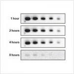
 | Western blot analysis of various lysates using INCENP Rabbit pAb (A0622) at 1:1000 dilution. Secondary antibody: HRP-conjugated Goat anti-Rabbit IgG (H+L) (AS014) at 1:10000 dilution. Lysates/proteins: 25μg per lane. Blocking buffer: 3% nonfat dry milk in TBST. Detection: ECL Basic Kit (RM00020). Exposure time: 30s. |
You may also be interested in:


