Your shopping cart is empty!
| Reactivity: | Human, Mouse, Rat |
| Applications: | WB, IF/IC, ELISA |
| Host Species: | Rabbit |
| Isotype: | IgG |
| Clonality: | Polyclonal antibody |
| Gene Name: | Ret proto-oncogene |
| Gene Symbol: | RET |
| Synonyms: | PTC; MTC1; HSCR1; MEN2A; MEN2B; CDHF12; CDHR16; RET-ELE1; Ret |
| Gene ID: | 5979 |
| UniProt ID: | P07949 |
| Immunogen: | A synthetic peptide corresponding to a sequence within amino acids 1000-1100 of human Ret (NP_066124.1). |
| Dilution: | WB 1:500-1:1000; IF/IC 1:50-1:200 |
| Purification Method: | Affinity purification |
| Concentration: | 2.25 mg/ml |
| Buffer: | PBS with 0.05% proclin300, 50% glycerol, pH7.3. |
| Storage: | Store at -20°C. Avoid freeze / thaw cycles. |
| Documents: | Manual-RET antibody |
Background
This gene encodes a transmembrane receptor and member of the tyrosine protein kinase family of proteins. Binding of ligands such as GDNF (glial cell-line derived neurotrophic factor) and other related proteins to the encoded receptor stimulates receptor dimerization and activation of downstream signaling pathways that play a role in cell differentiation, growth, migration and survival. The encoded receptor is important in development of the nervous system, and the development of organs and tissues derived from the neural crest. This proto-oncogene can undergo oncogenic activation through both cytogenetic rearrangement and activating point mutations. Mutations in this gene are associated with Hirschsprung disease and central hypoventilation syndrome and have been identified in patients with renal agenesis.
Images
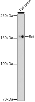 | Western blot analysis of lysates from Rat brain, using Ret Rabbit pAb (A0018) at 1:1000 dilution. Secondary antibody: HRP-conjugated Goat anti-Rabbit IgG (H+L) (AS014) at 1:10000 dilution. Lysates/proteins: 25μg per lane. Blocking buffer: 3% nonfat dry milk in TBST. Detection: ECL Enhanced Kit (RM00021). Exposure time: 180s. |
 | Immunofluorescence analysis of Neuro-2a cells using Ret Rabbit pAb (A0018) at dilution of 1:50 (40x lens). Secondary antibody: Cy3-conjugated Goat anti-Rabbit IgG (H+L) (AS007) at 1:500 dilution. Blue: DAPI for nuclear staining. |
 | Immunofluorescence analysis of SH-SY5Y cells using Ret Rabbit pAb (A0018) at dilution of 1:50 (40x lens). Secondary antibody: Cy3-conjugated Goat anti-Rabbit IgG (H+L) (AS007) at 1:500 dilution. Blue: DAPI for nuclear staining. |
You may also be interested in:
