Your shopping cart is empty!
The LiFluor™ Animal Cell Viability Kit offers a dual-color fluorescence staining method for distinguishing live and dead cells, utilizing two probes that assess two key parameters of cell viability: intracellular esterase activity and plasma membrane integrity. This versatile kit is compatible with a range of fluorescence detection systems, including fluorescence microscopes, multiwell plate scanners, flow cytometers, and other similar devices. The assay principles are broadly applicable to the majority of eukaryotic cell types, such as adherent cells and certain tissues, but are not suitable for bacteria or yeast. It is recognized for being faster, more cost-effective, safer, and a more sensitive indicator of cytotoxic events compared to alternative methods.
Live cells are identified by their ubiquitous intracellular esterase activity, which is measured by the enzymatic conversion of the nearly nonfluorescent cell-permeant calcein AM into the intensely fluorescent calcein. The polyanionic dye calcein is efficiently retained within live cells, resulting in a bright, uniform green fluorescence (Excitation/Emission ~495 nm/~520 nm). In contrast, propidium iodide (PI) penetrates cells with compromised membranes and experiences a 30-fold increase in fluorescence upon binding to nucleic acids, leading to a vivid red fluorescence in dead cells (Excitation/Emission ~528 nm/~617 nm). Live cells, with their intact plasma membranes, exclude PI, further enhancing the kit's ability to differentiate between live and dead cells.
Key Features
• Simplicity: Reagents are simultaneously added, no wash steps are required.
• Specificity and reliability: Distinct color for live and dead cell.
• Versatility: Suitable for fluorescence microscope, flow cytometer or microplate reader.
Specifications
1. Platform: Fluorescence Microscopy, Flow Cytometry
2. Detection Method: Fluorescent
3. Ex/Em: Calcein: 494/517; PI: 535/617 nm
Applications
Cell viability assay
Components
1. Calcein AM: 100 µl
2. Propidium Iodide: 100 µll
Storage
Store at -20°C and protect from light.
Case Study
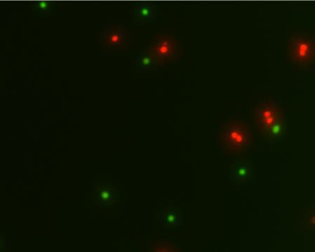
Live and dead Jurkat cells stained with LiFluor™ Animal Cell Viability Kit. Live cells fluoresce a bright green, whereas dead cells fluoresce red.
