PE Rabbit anti-Human CD100/SEMA4D mAb (100 T)
| Reactivity: | Human |
| Applications: | FC |
| Host Species: | Rabbit |
| Isotype: | IgG |
| Clonality: | Monoclonal Antibody |
| Conjugation: | PE. Ex:565nm. Em:574nm. |
| Gene Name: | semaphorin 4D |
| Gene Symbol: | SEMA4D |
| Synonyms: | A8; GR3; BB18; CD100; COLL4; SEMAJ; coll-4; C9orf164; M-sema-G |
| Gene ID: | 10507 |
| UniProt ID: | Q92854 |
| Immunogen: | Recombinant fusion protein containing a sequence corresponding to amino acids 22-734 of human Semaphorin-4D/CD100(NP_006369.3). |
| Dilution: | FC,5 μl per 10^6 cells in 100 μl volume |
| Purification Method: | Affinity purification |
| Concentration: | 0.04 mg/ml |
| Buffer: | PBS with 0.09% Sodium azide,0.2% BSA, pH7.3. |
| Storage: | Store at -20°C. Avoid freeze / thaw cycles. |
| Documents: | Manual-SEMA4D Antibody |
Background
Plexin-B1 is related to axon guidance in the central nervous system. The Sema4D function is also widely detected in the immune system and was found to be the first semaphorin expressed on the surface of many types of immune cells. In the immune system, CD72 is a low-affinity receptor for Sema4D, and studies have shown that Sema4D can not only regulate T cell activation, but also participate in the regulation of B cell survival and differentiation. In the immune system, soluble fragments containing extracellular domains produced by proteolytic cleavage can regulate many physiological functions of Sema4D. Sema4D is also associated with tumorigenesis because studies have confirmed that it is overexpressed in various types of solid tumor cells. To some extent, the role of Sema4D in tumorigenesis is related to its ability to cause tumor angiogenesis, cell invasion, and immunosuppression by enhancing bone marrow-derived suppressor cell function.
Images
 | Flow cytometry: 1×10^6 PC-3 cells (Low Expression,left) and MOLT-4 cells (right) were surface-stained with PE Rabbit anti-Human CD100/SEMA4D mAb (A26506,5 μl/Test,orange line) or PE Rabbit IgG isotype control (A24172,5 μl/Test,blue line). Non-fluorescently stained cells were used as blank control (red line). |
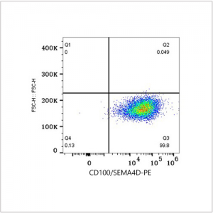
 | Flow cytometry: 1×10^6 MOLT-4 cells were surface-stained with PE Rabbit IgG isotype control (A24172,5 μl/Test,left) or PE Rabbit anti-Human CD100/SEMA4D mAb (A26506,5 μl/Test,right). |
You may also be interested in:


