Your shopping cart is empty!
")
KD-Validated EGFR Rabbit PolymAb® (20 μl)
| Reactivity: | Human |
| Applications: | WB, IF/IC, ELISA |
| Host Species: | Rabbit |
| Isotype: | IgG |
| Clonality: | Monoclonal antibody |
| Gene Name: | Epidermal growth factor receptor |
| Gene Symbol: | EGFR |
| Synonyms: | EGFR; ERBB; ERBB1; HER1; NISBD2; PIG61; mENA; epidermal growth factor receptor |
| Gene ID: | 1956 |
| UniProt ID: | P00533 |
| Clone ID: | 8U3O8 |
| Immunogen: | Recombinant protein of human EGFR. |
| Dilution: | WB 1:500-1:2000; IF/IC 1:50-1:200 |
| Purification Method: | Affinity purification |
| Concentration: | 1.09 mg/mL |
| Buffer: | PBS with 0.05% proclin300, 0.05% BSA, 50% glycerol, pH7.3. |
| Storage: | Store at -20°C. Avoid freeze / thaw cycles. |
| Documents: | Manual-EGFR monoclonal antibody |
Background
The protein encoded by this gene is a transmembrane glycoprotein that is a member of the protein kinase superfamily. This protein is a receptor for members of the epidermal growth factor family. EGFR is a cell surface protein that binds to epidermal growth factor. Binding of the protein to a ligand induces receptor dimerization and tyrosine autophosphorylation and leads to cell proliferation. Mutations in this gene are associated with lung cancer.
Images
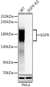 | Western blot analysis of lysates from wild type (WT) and EGFR knockdown (KD) HeLa cells, using [KD Validated] EGFR Rabbit PolymAb® (A24388PM) at1:1000 dilution. Secondary antibody: HRP-conjugated Goat anti-Rabbit IgG (H+L) (AS014) at 1:10000 dilution. Lysates/proteins: 25μg per lane. Blocking buffer: 3% nonfat dry milk in TBST. Detection: ECL Basic Kit (RM00020). Exposure time: 45s. |
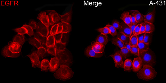 | Immunofluorescence analysis of A-431 cells using [KD Validated] EGFR Rabbit PolymAb® (A24388PM) at dilution of 1:100 (40x lens). Secondary antibody: Cy3-conjugated Goat anti-Rabbit IgG (H+L) (AS007) at 1:500 dilution. Blue: DAPI for nuclear staining. |
You may also be interested in: