APC Rabbit anti-Human CD1B mAb (100 T)
| Reactivity: | Human |
| Applications: | FC |
| Host Species: | Rabbit |
| Isotype: | IgG |
| Clonality: | Monoclonal Antibody |
| Conjugation: | APC. Ex:650nm. Em:660nm. |
| Gene Name: | CD1b molecule |
| Gene Symbol: | CD1B |
| Synonyms: | R1; CD1; CD1A |
| Gene ID: | 910 |
| UniProt ID: | P29016 |
| Immunogen: | Recombinant fusion protein containing a sequence corresponding to amino acids 18-303 of human CD1B (NP_001755.1). |
| Dilution: | FC,5 μl per 10^6 cells in 100 μl volume |
| Purification Method: | Affinity purification |
| Concentration: | 0.04 mg/ml |
| Buffer: | PBS with 0.09% Sodium azide,0.2% BSA, pH7.3. |
| Storage: | Store at -20°C. Avoid freeze / thaw cycles. |
| Documents: | Manual-CD1B Antibody |
Background
This gene encodes a member of the CD1 family of transmembrane glycoproteins, which are structurally related to the major histocompatibility complex (MHC) proteins and form heterodimers with beta-2-microglobulin. The CD1 proteins mediate the presentation of primarily lipid and glycolipid antigens of self or microbial origin to T cells. The human genome contains five CD1 family genes organized in a cluster on chromosome 1. The CD1 family members are thought to differ in their cellular localization and specificity for particular lipid ligands. The protein encoded by this gene localizes to late endosomes and lysosomes via a tyrosine-based motif in the cytoplasmic tail, and requires vesicular acidification to bind lipid antigens.
Images
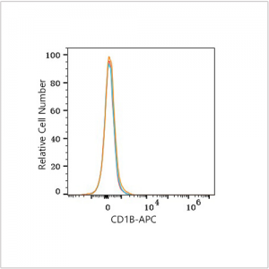
 | Flow cytometry: 1×10^6 K-562 cells (negative control,left) and MOLT-4 cells (right) were surface-stained with APC Rabbit anti-Human CD1B mAb (A26132,5 μl/Test,orange line) or APC Rabbit IgG isotype control (A24173,5 μl/Test,blue line). Non-fluorescently stained cells were used as blank control (red line). |
 | Flow cytometry: 1×10^6 MOLT-4 cells were surface-stained with APC Rabbit IgG isotype control (A24173,5 μl/Test,left) or APC Rabbit anti-Human CD1B mAb (A26132,5 μl/Test,right). |
You may also be interested in:


