PE Rabbit anti-Human CD146 mAb (100 T)
| Reactivity: | Human |
| Applications: | FC |
| Host Species: | Rabbit |
| Isotype: | IgG |
| Clonality: | Monoclonal Antibody |
| Conjugation: | PE. Ex:565nm. Em:574nm. |
| Gene Name: | melanoma cell adhesion molecule |
| Gene Symbol: | MCAM |
| Synonyms: | CD146; MUC18; HEMCAM; METCAM; MelCAM |
| Gene ID: | 4162 |
| UniProt ID: | P43121 |
| Clone ID: | 7P4C2 |
| Immunogen: | Recombinant fusion protein containing a sequence corresponding to amino acids 24-559 of human CD146 (NP_006491.2). |
| Dilution: | FC,5 μl per 10^6 cells in 100 μl volume |
| Purification Method: | Affinity purification |
| Concentration: | 0.01 mg/mL |
| Buffer: | PBS with 0.03% proclin300,0.2% BSA, pH7.3. |
| Storage: | Store at -20°C. Avoid freeze / thaw cycles. |
| Documents: | Manual-MCAM Antibody |
Background
Melanoma cell adhesion molecule (MCAM, MUC18, CD146) is an immunoglobulin superfamily member originally described as a cell surface adhesion protein and marker of the progression and metastasis of melanoma . Expression of MCAM protein is seen in vascular endothelial cells, activated T lymphocytes, smooth muscle, and bone marrow stromal cells. Research studies demonstrate increased MCAM expression in endothelial cells from angiogenesis-related disorders, including inflammatory bowel disease, Crohn’s disease, rheumatoid arthritis, tumors, and chronic renal failure . MCAM-expressing human mesenchymal stromal cells (hMSC) in the hematopoietic microenvironment are responsible for maintaining the self-renewal of hematopoietic stem and progenitor cells (HSPC) through direct contact between hMSC and HSPC. Related studies suggest that activation of the Notch signaling pathway may also, in part, play a role in HSPC maintenance . Additional research indicates that MCAM may play a role in multiple sclerosis, an autoimmune inflammatory disease that affects central nervous system neurons. Endothelial MCAM within the blood-brain barrier act as adhesion receptors that permit lymphocytes to transmigrate across the barrier and produce the inflammatory lesions that characterize the disorder .
Images
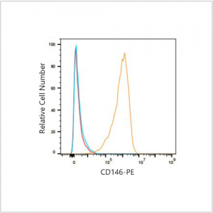
 | Flow cytometry: 1×10^6 MCF7 cells(negative control,left) and HUVEC(right) were surface-stained with PE Rabbit anti-Human CD146 mAb(A23748,5 μl/Test,orange line) or PE Rabbit IgG isotype control (5 μl/Test,blue line).Non-fluorescently stained cells were used as blank control (red line). |
 | Flow cytometry:1×10^6 HUVEC cells were surface-stained with PE Rabbit IgG isotype control (5 μl/Test,left) or PE Rabbit anti-Human CD146 mAb(A23748,5 μl/Test,right). |
You may also be interested in:


