| Reactivity: | Human |
| Applications: | WB, ELISA |
| Host Species: | Rabbit |
| Isotype: | IgG |
| Clonality: | Polyclonal antibody |
| Gene Name: | interleukin 17A |
| Gene Symbol: | IL17A |
| Synonyms: | IL17; CTLA8; IL-17; ILA17; CTLA-8; IL-17A; IL17A |
| Gene ID: | 3605 |
| UniProt ID: | Q16552 |
| Immunogen: | Recombinant fusion protein containing a sequence corresponding to amino acids 24-155 of human IL17A (NP_002181.1). |
| Dilution: | WB 1:500-1:1000; IF,1:50-1:200 |
| Purification Method: | Affinity purification |
| Concentration: | 1.87 mg/ml |
| Buffer: | PBS with 0.05% proclin300, 50% glycerol, pH7.3. |
| Storage: | Store at -20°C. Avoid freeze / thaw cycles. |
| Documents: | Manual-IL17A antibody |
Background
This gene is a member of the IL-17 receptor family which includes five members (IL-17RA-E) and the encoded protein is a proinflammatory cytokine produced by activated T cells. IL-17A-mediated downstream pathways induce the production of inflammatory molecules, chemokines, antimicrobial peptides, and remodeling proteins. The encoded protein elicits crucial impacts on host defense, cell trafficking, immune modulation, and tissue repair, with a key role in the induction of innate immune defenses. This cytokine stimulates non-hematopoietic cells and promotes chemokine production thereby attracting myeloid cells to inflammatory sites. This cytokine also regulates the activities of NF-kappaB and mitogen-activated protein kinases and can stimulate the expression of IL6 and cyclooxygenase-2 (PTGS2/COX-2), as well as enhance the production of nitric oxide (NO). IL-17A plays a pivotal role in various infectious diseases, inflammatory and autoimmune disorders, and cancer. High levels of this cytokine are associated with several chronic inflammatory diseases including rheumatoid arthritis, psoriasis and multiple sclerosis. The lung damage induced by the severe acute respiratory syndrome coronavirus 2 (SARS-CoV-2) is to a large extent, a result of the inflammatory response promoted by cytokines such as IL17A.
Images
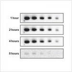
 | Western blot analysis of various lysates, using IL17A Rabbit pAb (A0688) at 1:500 dilution. Secondary antibody: HRP-conjugated Goat anti-Rabbit IgG (H+L) (AS014) at 1:10000 dilution. Lysates/proteins: 25μg per lane. Blocking buffer: 3% nonfat dry milk in TBST. Detection: ECL Basic Kit (RM00020). Exposure time: 30s. |
You may also be interested in:


