Your shopping cart is empty!
")
| Reactivity: | Human, Mouse |
| Applications: | WB, IHC, ELISA |
| Host Species: | Rabbit |
| Isotype: | IgG |
| Clonality: | Monoclonal antibody |
| Gene Name: | BCL2 antagonist/killer 1 |
| Gene Symbol: | BAK1 |
| Synonyms: | BAK; CDN1; BCL2L7; BAK-LIKE; Bak |
| Gene ID: | 578 |
| UniProt ID: | Q16611 |
| Clone ID: | 7Q3T8 |
| Immunogen: | Recombinant fusion protein containing a sequence corresponding to amino acids 11-82 of human Bak (NP_001179.1). |
| Dilution: | WB 1:1000-1:5000; IHC 1:1000-1:4000 |
| Purification Method: | Affinity purification |
| Concentration: | 0.09 mg/ml |
| Buffer: | PBS with 0.05% proclin300, 0.05% BSA, 50% glycerol, pH7.3. |
| Storage: | Store at -20°C. Avoid freeze / thaw cycles. |
| Documents: | Manual-BAK1 monoclonal antibody |
Background
The protein encoded by the gene BAK1 belongs to the BCL2 protein family. BCL2 family members form oligomers or heterodimers and act as anti- or pro-apoptotic regulators that are involved in a wide variety of cellular activities. This protein localizes to mitochondria, and functions to induce apoptosis. It interacts with and accelerates the opening of the mitochondrial voltage-dependent anion channel, which leads to a loss in membrane potential and the release of cytochrome c. This protein also interacts with the tumor suppressor P53 after exposure to cell stress.
Images
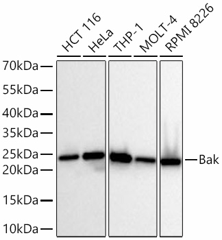 | Western blot analysis of various lysates, using [KD Validated] Bak Rabbit mAb (A23523) at 1:2000 dilution. Secondary antibody: HRP Goat Anti-Rabbit IgG (H+L) (AS014) at 1:10000 dilution. Lysates/proteins: 25μg per lane. Blocking buffer: 3% nonfat dry milk in TBST. Detection: ECL Basic Kit (RM00020). Exposure time: 10s. |
 | Western blot analysis of various lysates, using [KD Validated] Bak Rabbit mAb (A23523) at 1:2000 dilution. Secondary antibody: HRP-conjugated Goat anti-Rabbit IgG (H+L) (AS014) at 1:10000 dilution. Lysates/proteins: 25μg per lane. Blocking buffer: 3% nonfat dry milk in TBST. Detection: ECL Basic Kit (RM00020). Exposure time: 10s. |
 | Western blot analysis of lysates from wild type (WT) and Bak knockdown (KD) 293T cells using [KD Validated] Bak Rabbit mAb (A23523) at 1:1000 dilution incubated overnight at 4℃. Secondary antibody: HRP-conjugated Goat anti-Rabbit IgG (H+L) (AS014) at 1:10000 dilution. Lysates/proteins: 25 μg per lane. Blocking buffer: 3% nonfat dry milk in TBST. Detection: ECL Basic Kit (RM00020). Exposure time: 20s. |
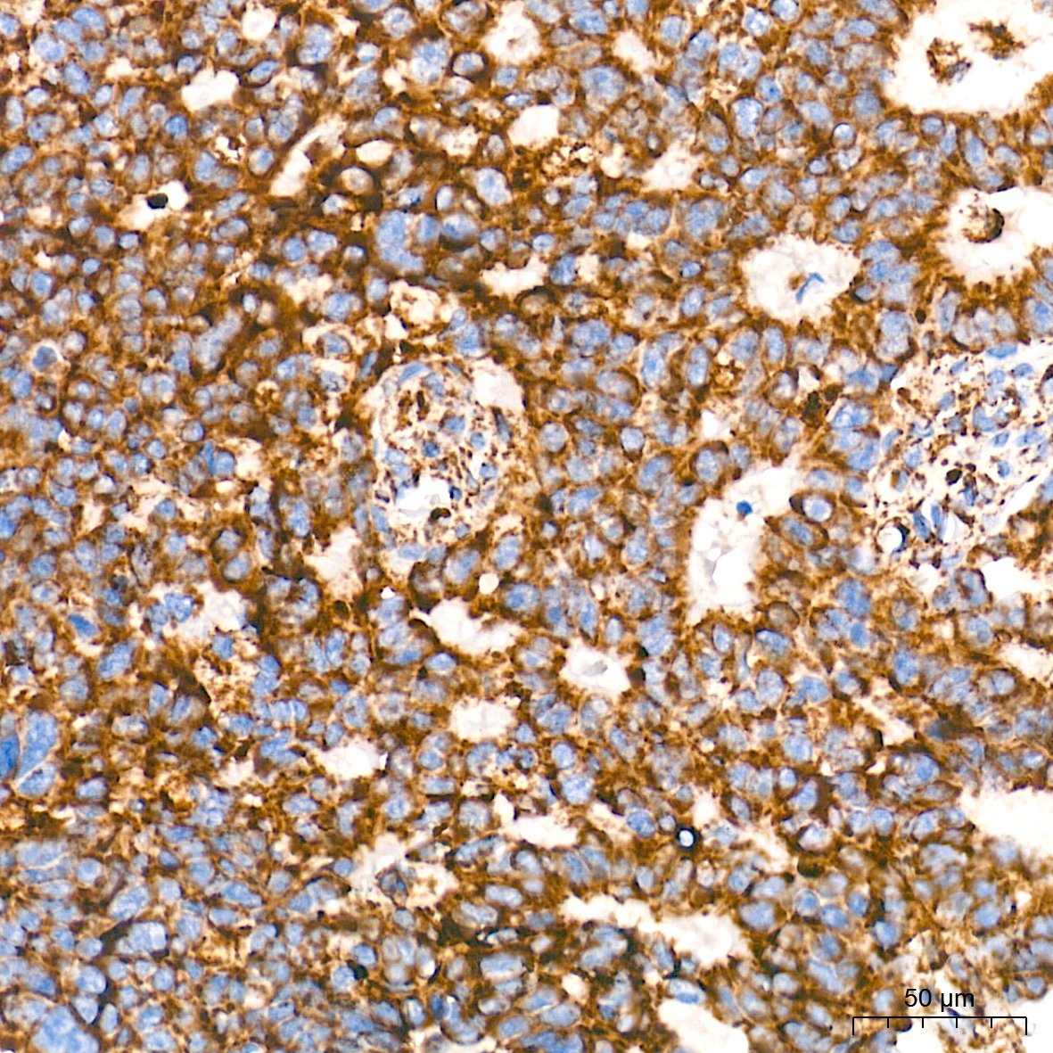
 | Immunohistochemistry analysis of paraffin-embedded Human colon carcinoma tissue using [KD Validated] Bak Rabbit mAb (A23523) at a dilution of 1:200 (40x lens). High pressure antigen retrieval performed with 0.01M Tris-EDTA Buffer (pH 9.0) prior to IHC staining. |
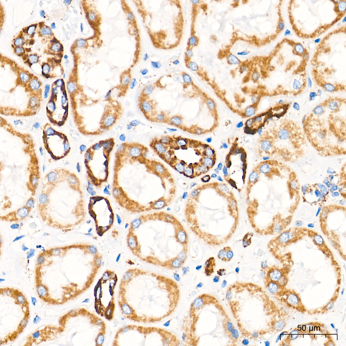
 | Immunohistochemistry analysis of paraffin-embedded Human kidney tissue using [KD Validated] Bak Rabbit mAb (A23523) at a dilution of 1:200 (40x lens). High pressure antigen retrieval performed with 0.01M Tris-EDTA Buffer (pH 9.0) prior to IHC staining. |
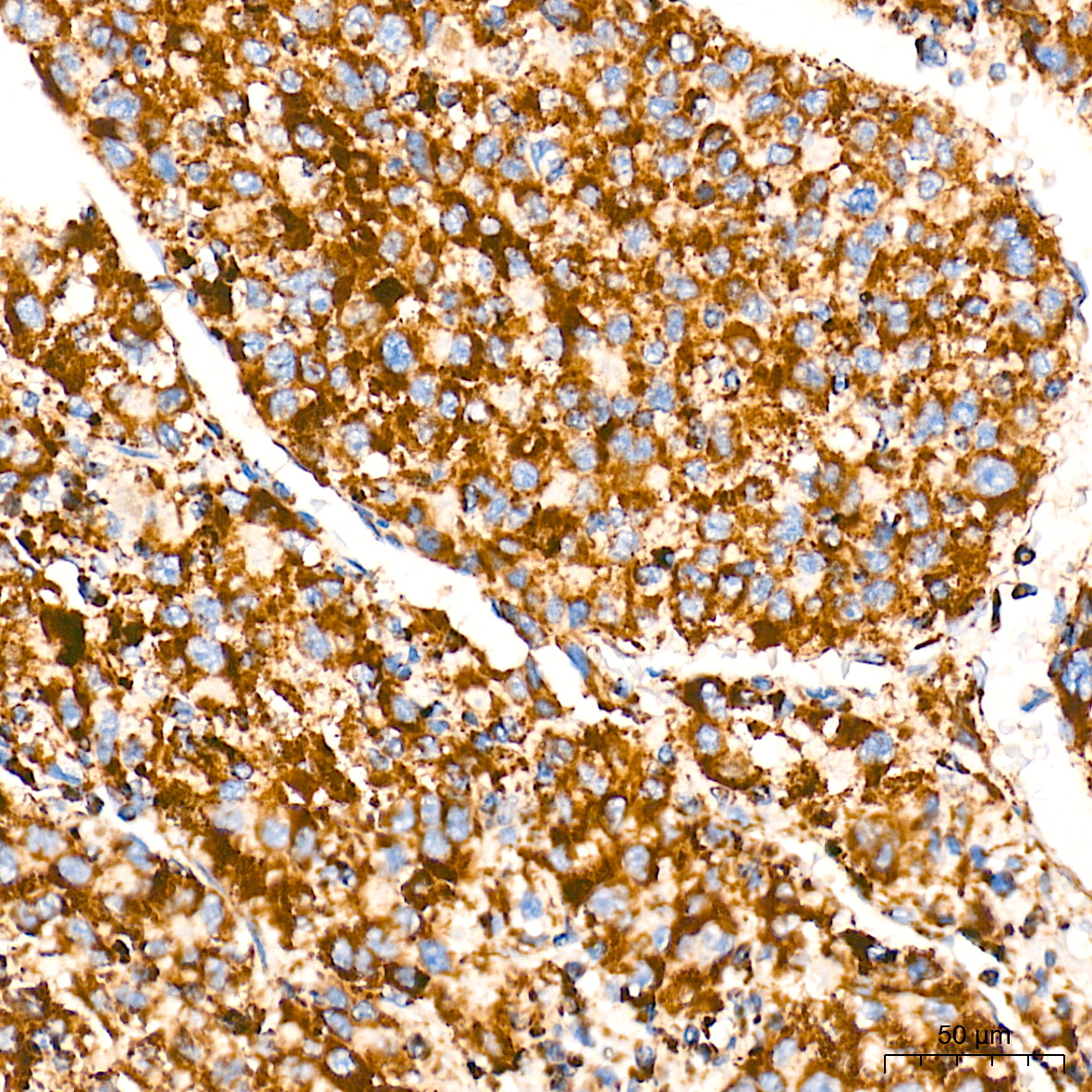
 | Immunohistochemistry analysis of paraffin-embedded Human liver cancer tissue using [KD Validated] Bak Rabbit mAb (A23523) at a dilution of 1:200 (40x lens). High pressure antigen retrieval performed with 0.01M Tris-EDTA Buffer (pH 9.0) prior to IHC staining. |
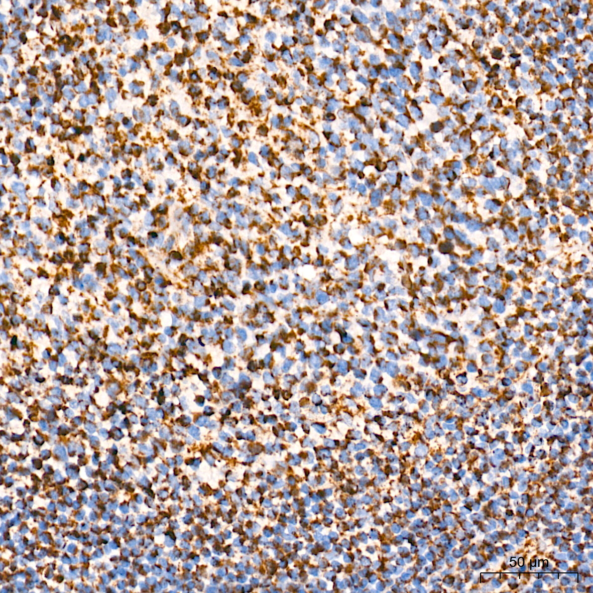
 | Immunohistochemistry analysis of paraffin-embedded Human tonsil tissue using [KD Validated] Bak Rabbit mAb (A23523) at a dilution of 1:200 (40x lens). High pressure antigen retrieval performed with 0.01M Tris-EDTA Buffer (pH 9.0) prior to IHC staining. |
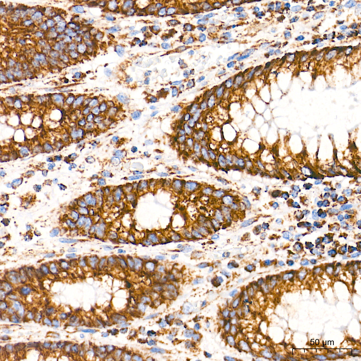
 | Immunohistochemistry analysis of paraffin-embedded Human colon tissue using [KD Validated] Bak Rabbit mAb (A23523) at a dilution of 1:200 (40x lens). High pressure antigen retrieval performed with 0.01M Tris-EDTA Buffer (pH 9.0) prior to IHC staining. |
You may also be interested in:

