Your shopping cart is empty!
")
| Reactivity: | Human, Mouse, Rat |
| Applications: | WB,IHC,IF/IC,ELISA |
| Host Species: | Rabbit |
| Isotype: | IgG |
| Clonality: | Monoclonal antibody |
| Gene Name: | sphingosine-1-phosphate lyase 1 |
| Gene Symbol: | SGPL1 |
| Synonyms: | SPL; S1PL; NPHS14 |
| Gene ID: | 8879 |
| UniProt ID: | O95470 |
| Immunogen: | A synthetic peptide corresponding to a sequence within amino acids 301-400 of human SGPL1 (NP_003892.2). |
| Dilution: | WB 1:7500-1:30000; IHC 1:100-1:400; IF/IC 1:200-1:800 |
| Purification Method: | Affinity purification |
| Concentration: | 1.73 mg/ml |
| Buffer: | PBS with 0.09% Sodium azide, 0.05% BSA, 50% glycerol, pH7.3. |
| Storage: | Store at -20°C. Avoid freeze / thaw cycles. |
| Documents: | Manual-SGPL1 monoclonal antibody |
Background
Enables sphinganine-1-phosphate aldolase activity. Involved in apoptotic signaling pathway; fatty acid metabolic process; and sphingolipid metabolic process. Located in endoplasmic reticulum. Implicated in nephrotic syndrome type 14.
Images
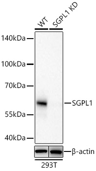 | Western blot analysis of lysates from wild type (WT) and SGPL1 knockout (KD) 293T cells using [KD Validated] SGPL1 Rabbit mAb (A26855) at 1:15000 dilution incubated overnight at 4℃. Secondary antibody: HRP-conjugated Goat anti-Rabbit IgG (H+L) (AS014) at 1:10000 dilution. Lysates/proteins: 25 μg per lane. Blocking buffer: 3% nonfat dry milk in TBST. Detection: ECL Basic Kit (RM00020). Exposure time: 45s. |
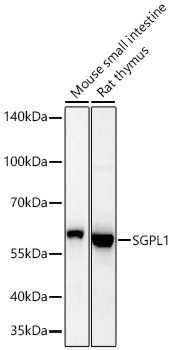 | Western blot analysis of various lysates using [KD Validated] SGPL1 Rabbit mAb (A26855) at 1:15000 dilution incubated overnight at 4℃. Secondary antibody: HRP-conjugated Goat anti-Rabbit IgG (H+L) (AS014) at 1:10000 dilution. Lysates/proteins: 25 μg per lane. Blocking buffer: 3% nonfat dry milk in TBST. Detection: ECL Basic Kit (RM00020). Exposure time: 45s. |
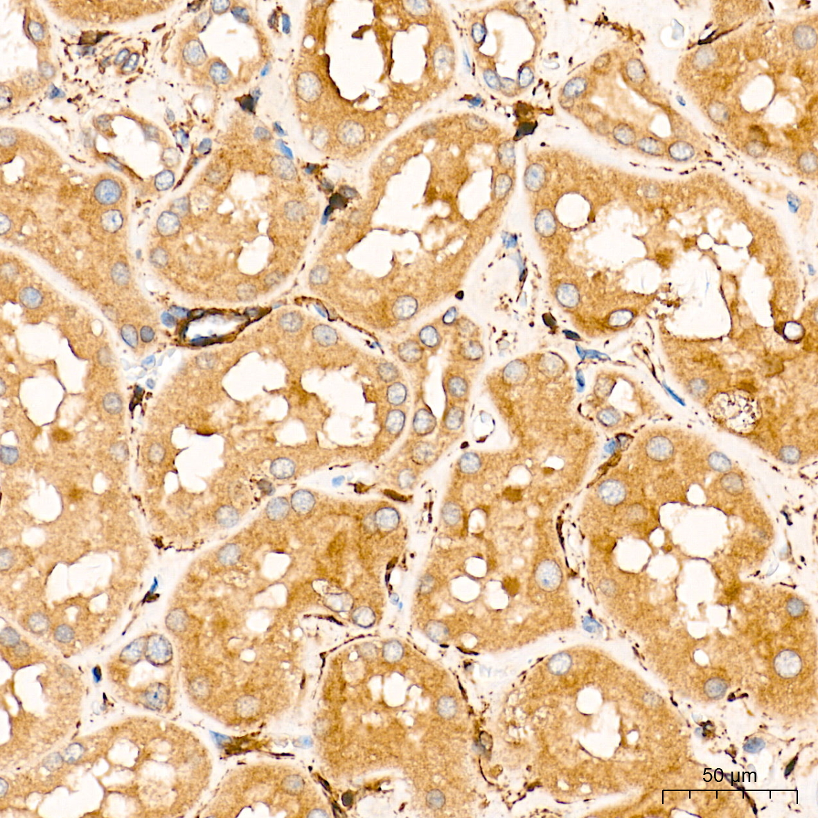 | Immunohistochemistry analysis of paraffin-embedded Human kidney tissue using [KD Validated] SGPL1 Rabbit mAb (A26855) at a dilution of 1:200 (40x lens). High pressure antigen retrieval performed with 0.01M Citrate Buffer (pH 6.0) prior to IHC staining. |
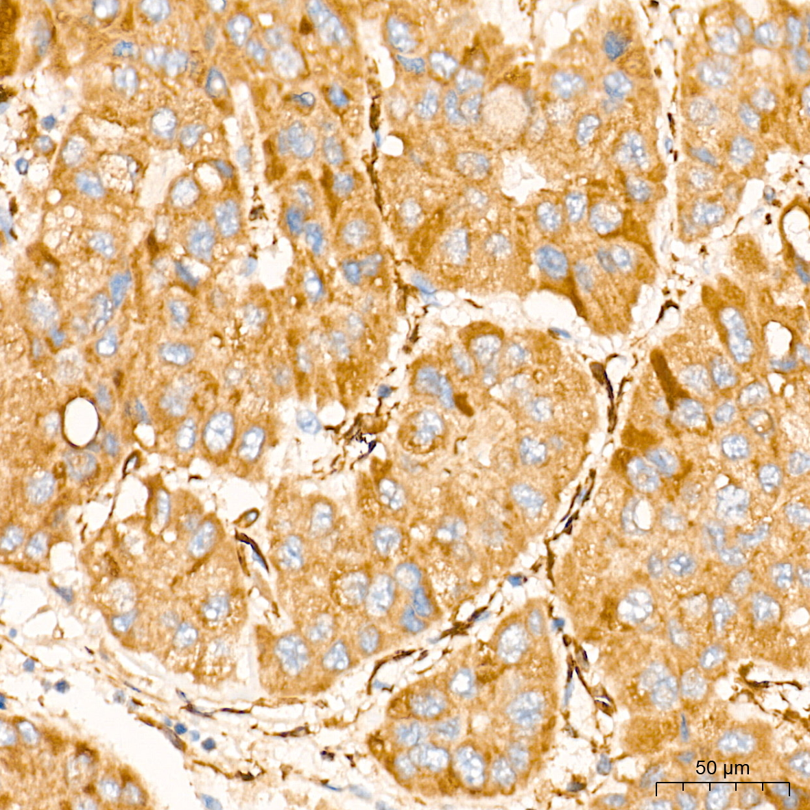 | Immunohistochemistry analysis of paraffin-embedded Human liver cancer tissue using [KD Validated] SGPL1 Rabbit mAb (A26855) at a dilution of 1:200 (40x lens). High pressure antigen retrieval performed with 0.01M Citrate Buffer (pH 6.0) prior to IHC staining. |
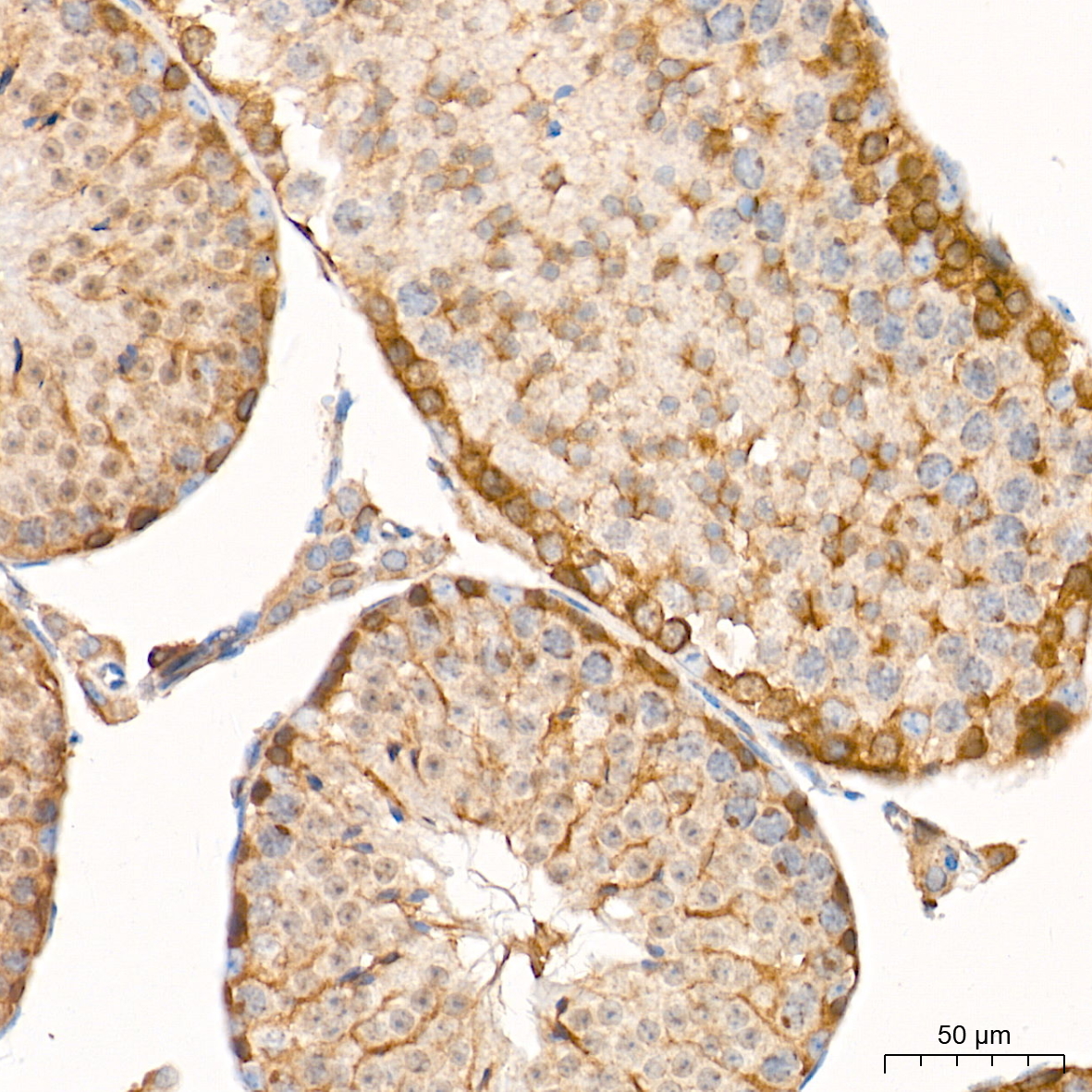 | Immunohistochemistry analysis of paraffin-embedded Mouse testis tissue using [KD Validated] SGPL1 Rabbit mAb (A26855) at a dilution of 1:200 (40x lens). High pressure antigen retrieval performed with 0.01M Citrate Buffer (pH 6.0) prior to IHC staining. |
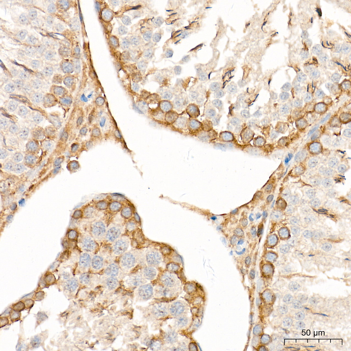 | Immunohistochemistry analysis of paraffin-embedded Rat testis tissue using [KD Validated] SGPL1 Rabbit mAb (A26855) at a dilution of 1:200 (40x lens). High pressure antigen retrieval performed with 0.01M Citrate Buffer (pH 6.0) prior to IHC staining. |
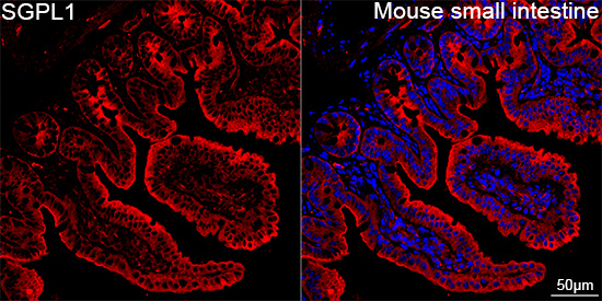 | Confocal imaging of paraffin-embedded Mouse small intesine tissue using [KD Validated] SGPL1 Rabbit mAb (A26855, dilution 1:200) followed by a further incubation with Cy3 Goat Anti-Rabbit IgG (H+L) (AS007, dilution 1:500) (Red). DAPI was used for nuclear staining (Blue). High pressure antigen retrieval performed with 0.01M Citrate Buffer(pH 6.0) prior to IF staining. Objective: 40x. |
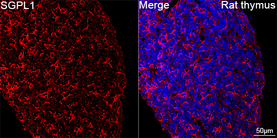 | Confocal imaging of paraffin-embedded Rat thymus tissue using [KD Validated] SGPL1 Rabbit mAb (A26855, dilution 1:200) followed by a further incubation with Cy3 Goat Anti-Rabbit IgG (H+L) (AS007, dilution 1:500) (Red). DAPI was used for nuclear staining (Blue). High pressure antigen retrieval performed with 0.01M Citrate Buffer(pH 6.0) prior to IF staining. Objective: 40x. |
You may also be interested in: