Your shopping cart is empty!
")
| Reactivity: | Human, Mouse, Rat |
| Applications: | WB, IHC, IF/IC, ELISA |
| Host Species: | Rabbit |
| Isotype: | IgG |
| Clonality: | Polyclonal antibody |
| Gene Name: | transcription factor EB |
| Gene Symbol: | TFEB |
| Synonyms: | TCFEB; BHLHE35; ALPHATFEB; TFEB |
| Gene ID: | 7942 |
| UniProt ID: | P19484 |
| Immunogen: | Recombinant fusion protein containing a sequence corresponding to amino acids 50-173 of human TFEB (NP_009093.1). |
| Dilution: | WB 1:500-1:2000; IHC 1:50-1:200; IF/IC 1:50-1:200 |
| Purification Method: | Affinity purification |
| Concentration: | 1.08 mg/mL |
| Buffer: | PBS with 0.02% sodium azide, 50% glycerol ,pH7.3. |
| Storage: | Store at -20°C. Avoid freeze / thaw cycles. |
| Documents: | Manual-TFEB polyclonal antibody |
Background
Enables DNA-binding transcription factor activity; enzyme binding activity; and transcription cis-regulatory region binding activity. Involved in several processes, including cellular response to amino acid starvation; lysosome localization; and positive regulation of autophagy. Located in cytosol; lysosomal membrane; and nucleoplasm.
Images
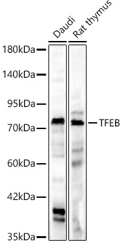 | Western blot analysis of various lysates, using [KD Validated] TFEB Rabbit pAb (A7311) at 1:1000 dilution. Secondary antibody: HRP-conjugated Goat anti-Rabbit IgG (H+L) (AS014) at 1:10000 dilution. Lysates/proteins: 25μg per lane. Blocking buffer: 3% nonfat dry milk in TBST. Detection: ECL Basic Kit (RM00020). Exposure time: 180s. |
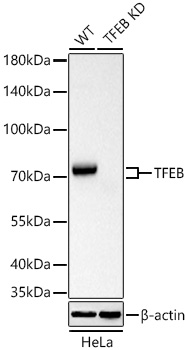 | Western blot analysis of lysates from wild type (WT) and TFEB knockdown (KD) HeLa cells using [KD Validated] TFEB Rabbit pAb (A7311) at 1:1000 dilution incubated overnight at 4℃. Secondary antibody: HRP-conjugated Goat anti-Rabbit IgG (H+L) (AS014) at 1:10000 dilution. Lysates/proteins: 25 μg per lane. Blocking buffer: 3% nonfat dry milk in TBST. Detection: ECL Basic Kit (RM00020). Exposure time: 10s. |
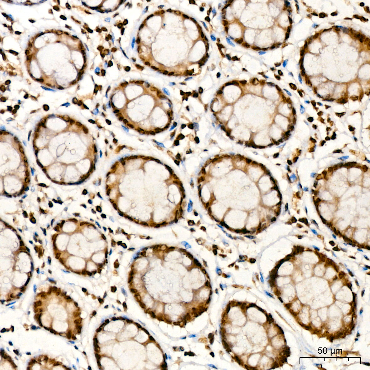 | Immunohistochemistry analysis of paraffin-embedded Human colon tissue using [KD Validated] TFEB Rabbit pAb (A7311) at a dilution of 1:100 (40x lens). High pressure antigen retrieval was performed with 0.01 M citrate buffer (pH 6.0) prior to IHC staining. |
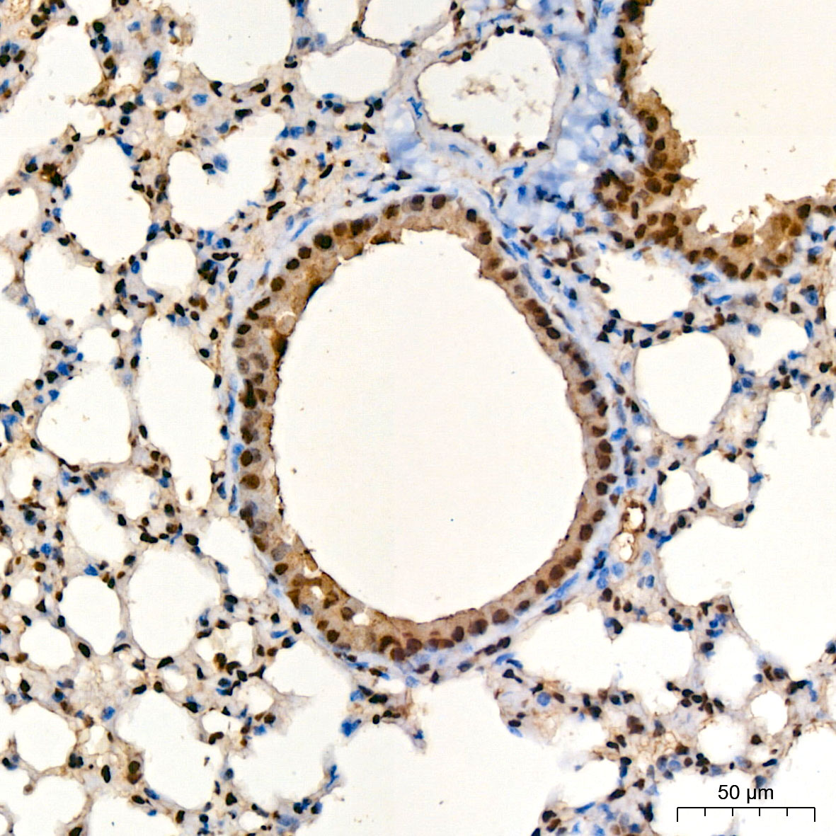 | Immunohistochemistry analysis of paraffin-embedded Mouse lung tissue using [KD Validated] TFEB Rabbit pAb (A7311) at a dilution of 1:100 (40x lens). High pressure antigen retrieval was performed with 0.01 M citrate buffer (pH 6.0) prior to IHC staining. |
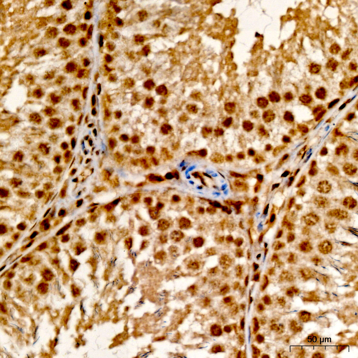 | Immunohistochemistry analysis of paraffin-embedded Rat testis tissue using [KD Validated] TFEB Rabbit pAb (A7311) at a dilution of 1:100 (40x lens). High pressure antigen retrieval was performed with 0.01 M citrate buffer (pH 6.0) prior to IHC staining. |
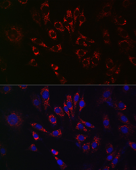 | Immunofluorescence analysis of NIH/3T3 cells using [KD Validated] TFEB Rabbit pAb (A7311) at dilution of 1:50 (40x lens). Secondary antibody: Cy3-conjugated Goat anti-Rabbit IgG (H+L) (AS007) at 1:500 dilution. Blue: DAPI for nuclear staining. |
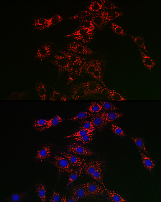 | Immunofluorescence analysis of PC-12 cells using [KD Validated] TFEB Rabbit pAb (A7311) at dilution of 1:50 (40x lens). Secondary antibody: Cy3-conjugated Goat anti-Rabbit IgG (H+L) (AS007) at 1:500 dilution. Blue: DAPI for nuclear staining. |
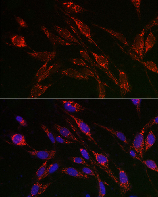 | Immunofluorescence analysis of U2OS cells using [KD Validated] TFEB Rabbit pAb (A7311) at dilution of 1:50 (40x lens). Secondary antibody: Cy3-conjugated Goat anti-Rabbit IgG (H+L) (AS007) at 1:500 dilution. Blue: DAPI for nuclear staining. |
You may also be interested in: