| Reactivity: | Human, Mouse, Rat |
| Applications: | WB, FC |
| Host Species: | Rabbit |
| Isotype: | IgG |
| Clonality: | Monoclonal antibody |
| Gene Name: | SMAD family member 3 |
| Gene Symbol: | SMAD3 |
| Synonyms: | LDS3; mad3; LDS1C; MADH3; JV15-2; hMAD-3; hSMAD3; HSPC193; HsT17436 |
| Gene ID: | 4088 |
| UniProt ID: | P84022 |
| Clone ID: | 25GB3215 |
| Immunogen: | A synthesized peptide derived from human Phospho-Smad3 (S423 + S425) |
| Dilution: | WB 1:4,000-1:20,000; FC 1:2,000 |
| Purification Method: | Affinity purified |
| Concentration: | Lot dependent |
| Buffer: | PBS with 0.02% sodium azide, 50% glycerol, pH7.3. |
| Storage: | Store at -20°C. Avoid freeze/thaw cycles. |
Background
The SMAD family of proteins are a group of intracellular signal transducer proteins similar to the gene products of the Drosophila gene 'mothers against decapentaplegic' (Mad) and the C. elegans gene Sma. The SMAD3 protein functions in the transforming growth factor-beta signaling pathway, and transmits signals from the cell surface to the nucleus, regulating gene activity and cell proliferation. This protein forms a complex with other SMAD proteins and binds DNA, functioning both as a transcription factor and tumor suppressor. Mutations in this gene are associated with aneurysms-osteoarthritis syndrome and Loeys-Dietz Syndrome 3.
Images
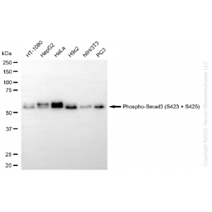
 | Western blotting analysis using anti-phospho-Smad3 (S423 + S425) antibody (Cat#71222). Total cell lysates (30 μg) from various cell lines were loaded and separated by SDS-PAGE. The blot was incubated with anti-phospho-Smad3 (S423 + S425) antibody (Cat#71222, 1:5,000) and HRP-conjugated goat anti rabbit secondary antibody (Cat#201, 1:20,000) respectively. Image was developed using FeQ™ ECL Substrate Kit (Cat#226). |
 | Western blotting analysis using anti-phospho-smad3 (S423 + S425) antibody (Cat#71222). Phospho-smad3 (S423 + S425) expression in wild-type (WT) and SMAD3 knockout (KO) HSHC cells with 20 μg of total cell lysates. Hsp90 α serves as a loading control. The blot was incubated with anti-phospho-smad3 (S423 + S425) antibody (Cat#71222, 1:5,000) and HRP-conjugated goat anti-rabbit secondary antibody (Cat#201, 1:20,000) respectively. Image was developed using NaQ™ ECL Substrate Kit (Cat#716). |
 | Flow cytometric analysis of Phospho-Smad3 (S423 + S425) expression in HAP-1 cells using anti-Phospho-Smad3 (S423 + S425) antibody (Cat#71222, 1:2,000). Green, isotype control; red, Phospho-Smad3 (S423 + S425). |
 | lmmunocytochemical staining of HepG2 cells with YAP1 antibody (Cat#71223, 1:1,000). Nuclei were stained blue with DAPl; YAP1 was stained magenta with Alexa Fluor® 647. lmages were taken using Leica stellaris 5. Protein abundance based on laser Intensity and smart gain: Medium. Scale bar, 20 μm. |
You may also be interested in:

