Your shopping cart is empty!
")
| Reactivity: | Human, Mouse, Rat |
| Applications: | WB, IF/IC, ELISA |
| Host Species: | Rabbit |
| Isotype: | IgG |
| Clonality: | Polyclonal antibody |
| Gene Name: | GIT ArfGAP 1 |
| Gene Symbol: | GIT1 |
| Synonyms: | p95-APP1; T1 |
| Gene ID: | 28964 |
| UniProt ID: | Q9Y2X7 |
| Immunogen: | Recombinant fusion protein containing a sequence corresponding to amino acids 460-640 of human GIT1 (NP_054749.2). |
| Dilution: | WB 1:200-1:2000; IF/IC 1:50-1:200 |
| Purification Method: | Affinity purification |
| Concentration: | 0.86 mg/ml |
| Buffer: | PBS with 0.01% thimerosal, 50% glycerol, pH7.3. |
| Storage: | Store at -20°C. Avoid freeze / thaw cycles. |
| Documents: | Manual-GIT1 polyclonal antibody |
Background
Enables gamma-tubulin binding activity. Involved in positive regulation of microtubule nucleation and regulation of cytokinesis. Located in several cellular components, including focal adhesion; microtubule cytoskeleton; and mitochondrion. Implicated in attention deficit hyperactivity disorder. Biomarker of Huntington's disease.
Images
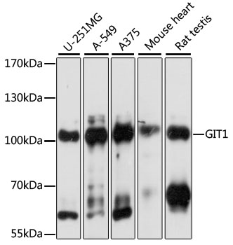 | Western blot analysis of various lysates using [KO Validated] GIT1 Rabbit pAb (A15437) at 1:1000 dilution. Secondary antibody: HRP-conjugated Goat anti-Rabbit IgG (H+L) (AS014) at 1:10000 dilution. Lysates/proteins: 25μg per lane. Blocking buffer: 3% nonfat dry milk in TBST. Detection: ECL Basic Kit (RM00020). Exposure time: 1s. |
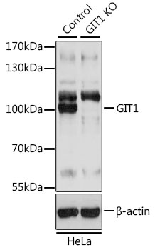 | Western blot analysis of lysates from wild type (WT) and GIT1 knockout (KO) HeLa cells, using [KO Validated] GIT1 Rabbit pAb (A15437) at 1:3000 dilution. Secondary antibody: HRP-conjugated Goat anti-Rabbit IgG (H+L) (AS014) at 1:10000 dilution. Lysates/proteins: 25μg per lane. Blocking buffer: 3% nonfat dry milk in TBST. Detection: ECL Basic Kit (RM00020). Exposure time: 1s. |
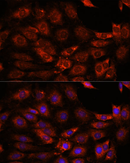 | Immunofluorescence analysis of C6 cells using [KO Validated] GIT1 Rabbit pAb (A15437) at dilution of 1:100. Secondary antibody: Cy3-conjugated Goat anti-Rabbit IgG (H+L) (AS007) at 1:500 dilution. Blue: DAPI for nuclear staining. |
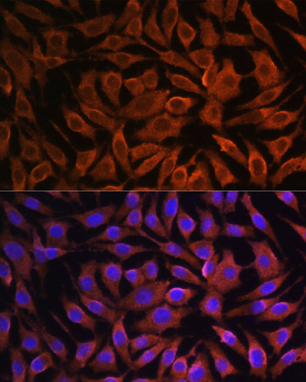 | Immunofluorescence analysis of L929 cells using [KO Validated] GIT1 Rabbit pAb (A15437) at dilution of 1:100. Secondary antibody: Cy3-conjugated Goat anti-Rabbit IgG (H+L) (AS007) at 1:500 dilution. Blue: DAPI for nuclear staining. |
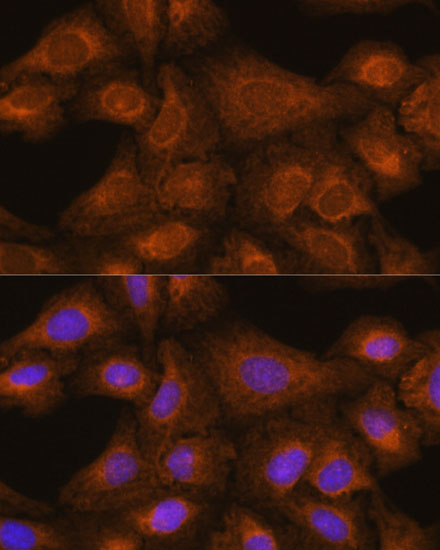 | Immunofluorescence analysis of U-2 OS cells using [KO Validated] GIT1 Rabbit pAb (A15437) at dilution of 1:100. Secondary antibody: Cy3-conjugated Goat anti-Rabbit IgG (H+L) (AS007) at 1:500 dilution. Blue: DAPI for nuclear staining. |
You may also be interested in:
