Your shopping cart is empty!
")
| Reactivity: | Human, Mouse, Rat |
| Applications: | WB, IHC, IF/IC, IP, ELISA |
| Host Species: | Rabbit |
| Isotype: | IgG |
| Clonality: | Monoclonal antibody |
| Gene Name: | Histone deacetylase 1 |
| Gene Symbol: | HDAC1 |
| Synonyms: | HD1; RPD3; KDAC1; GON-10; RPD3L1; C1 |
| Gene ID: | 3065 |
| UniProt ID: | Q13547 |
| Clone ID: | 2L6O7 |
| Immunogen: | A synthetic peptide corresponding to a sequence within amino acids 350-450 of human HDAC1 (Q13547). |
| Dilution: | WB 1:1000-1:4000; IHC 1:500-1:2000; IF/IC 1:100-1:2000 |
| Purification Method: | Affinity purification |
| Concentration: | 0.4 mg/mL |
| Buffer: | PBS with 0.02% sodium azide, 0.05% BSA, 50% glycerol, pH7.3. |
| Storage: | Store at -20°C. Avoid freeze / thaw cycles. |
| Documents: | Manual-HDAC1 monoclonal antibody |
Background
Histone acetylation and deacetylation, catalyzed by multisubunit complexes, play a key role in the regulation of eukaryotic gene expression. The protein encoded by this gene belongs to the histone deacetylase/acuc/apha family and is a component of the histone deacetylase complex. It also interacts with retinoblastoma tumor-suppressor protein and this complex is a key element in the control of cell proliferation and differentiation. Together with metastasis-associated protein-2, it deacetylates p53 and modulates its effect on cell growth and apoptosis.
Images
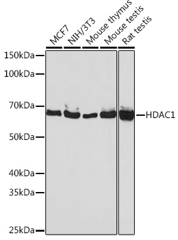 | Western blot analysis of various lysates using [KO Validated] HDAC1 Rabbit mAb (A19571) at 1:1000 dilution. Secondary antibody: HRP-conjugated Goat anti-Rabbit IgG (H+L) (AS014) at 1:10000 dilution. Lysates/proteins: 25μg per lane. Blocking buffer: 3% nonfat dry milk in TBST. Detection: ECL Basic Kit (RM00020). Exposure time: 30s. |
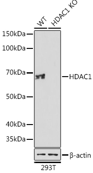 | Western blot analysis of lysates from wild type (WT) and HDAC1 knockout (KO) 293T cells, using [KO Validated] HDAC1 Rabbit mAb (A19571) at 1:1000 dilution. Secondary antibody: HRP-conjugated Goat anti-Rabbit IgG (H+L) (AS014) at 1:10000 dilution. Lysates/proteins: 25μg per lane. Blocking buffer: 3% nonfat dry milk in TBST. Detection: ECL Basic Kit (RM00020). Exposure time: 30s. |
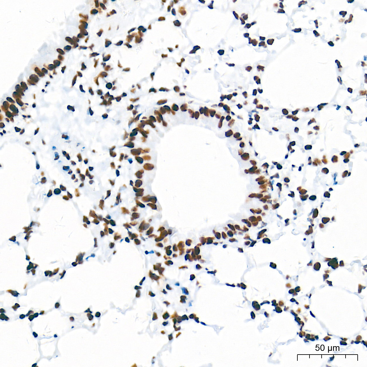 | Immunohistochemistry analysis of paraffin-embedded Mouse lung tissue using [KO Validated] HDAC1 Rabbit mAb (A19571) at a dilution of 1:2000 (40x lens). High pressure antigen retrieval performed with 0.01M Tris-EDTA Buffer (pH 9.0) prior to IHC staining. |
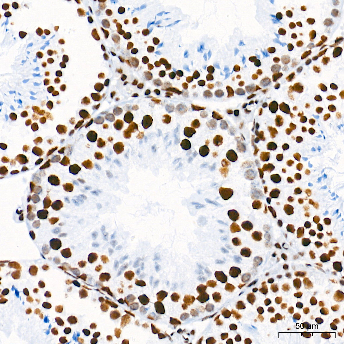 | Immunohistochemistry analysis of paraffin-embedded Mouse testis tissue using [KO Validated] HDAC1 Rabbit mAb (A19571) at a dilution of 1:2000 (40x lens). High pressure antigen retrieval performed with 0.01M Tris-EDTA Buffer (pH 9.0) prior to IHC staining. |
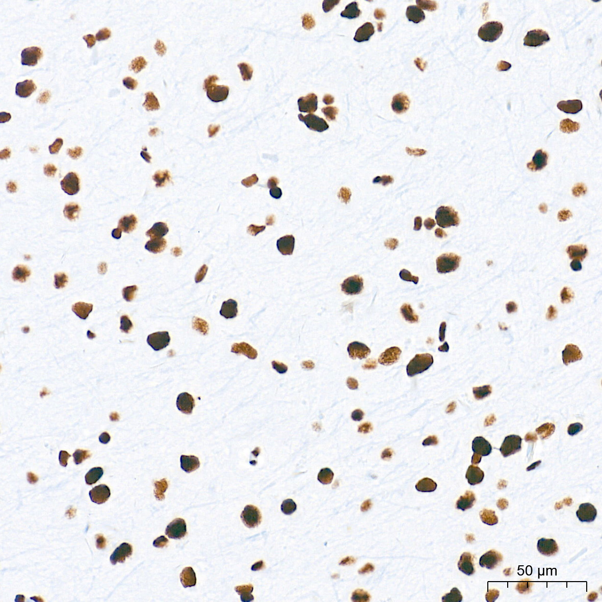 | Immunohistochemistry analysis of paraffin-embedded Rat brain tissue using [KO Validated] HDAC1 Rabbit mAb (A19571) at a dilution of 1:2000 (40x lens). High pressure antigen retrieval performed with 0.01M Tris-EDTA Buffer (pH 9.0) prior to IHC staining. |
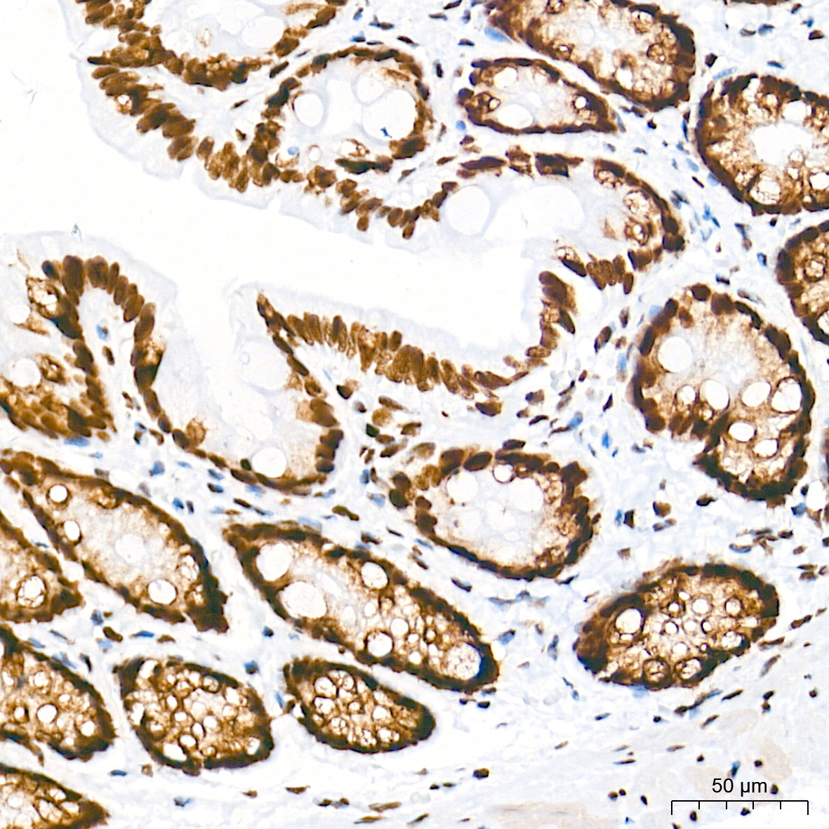 | Immunohistochemistry analysis of paraffin-embedded Rat colon tissue using [KO Validated] HDAC1 Rabbit mAb (A19571) at a dilution of 1:2000 (40x lens). High pressure antigen retrieval performed with 0.01M Tris-EDTA Buffer (pH 9.0) prior to IHC staining. |
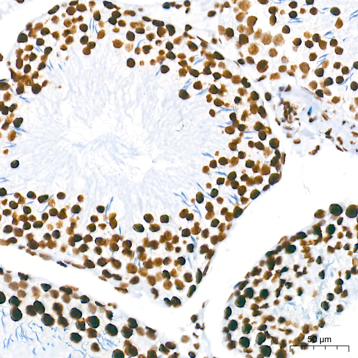 | Immunohistochemistry analysis of paraffin-embedded Rat testis tissue using [KO Validated] HDAC1 Rabbit mAb (A19571) at a dilution of 1:2000 (40x lens). High pressure antigen retrieval performed with 0.01M Tris-EDTA Buffer (pH 9.0) prior to IHC staining. |
 | Immunohistochemistry analysis of paraffin-embedded Mouse lung tissue using [KO Validated] HDAC1 Rabbit mAb (A19571) at a dilution of 1:2000 (40x lens). High pressure antigen retrieval performed with 0.01M Tris-EDTA Buffer (pH 9.0) prior to IHC staining. |
You may also be interested in: