Your shopping cart is empty!
 Rabbit pAb (50 µl)")
| Reactivity: | Human, Mouse, Rat, Monkey |
| Applications: | WB, IHC, IF/IC |
| Host Species: | Rabbit |
| Clonality: | Polyclonal |
| Full Name: | Rabbit anti-HSP90 beta (pS254) Antibody |
| Synonyms: | HSP84; HSPC2; HSPCB; D6S182; HSP90B |
| Immunogen: | KLH-conjugated synthetic peptide encompassing a sequence within the center region of human HSP90 beta (pS254). The exact sequence is proprietary. |
| Dilution: | WB 1:500-1:1000; IHC 1:50-1:100; IF/IC 1:50-1:200 |
| Gene ID (Human): | 3326 |
| Gene ID (Mouse): | 15516 |
| Gene ID (Rat): | 301252 |
| SwissProt (Human): | P08238 |
| SwissProt (Mouse): | P11499 |
| SwissProt (Rat): | P34058 |
| Purification Method: | The antibody was purified by immunogen affinity chromatography. |
| Concentration: | 1mg/mL |
| Buffer: | Liquid in 0.42% Potassium phosphate, 0.87% Sodium chloride, pH 7.3, 30% glycerol, and 0.01% sodium azide. |
| Storage: | Store at -20°C. Avoid repeated freeze / thaw cycles. |
| Documents: | Manual |
Images
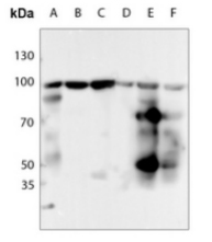
Western blot analysis of HSP90 beta (Phospho-S254) expression in HEK293T (A), Hela (B), U2OS (C), mouse testis (D), rat lung (E), rat testis (F) whole cell lysates. (Predicted band size: 83 kD; Observed band size: 90 kD)
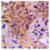
Immunohistochemical analysis of HSP90 beta (Phospho-S254) staining in human breast cancer formalin fixed paraffin embedded tissue section. The section was pre-treated using heat mediated antigen retrieval with sodium citrate buffer (pH 6.0). The section was then incubated with the antibody at room temperature and detected using an HRP conjugated compact polymer system. DAB was used as the chromogen. The section was then counterstained with haematoxylin and mounted with DPX.
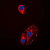
Immunofluorescent analysis of HSP90 beta (Phospho-S254) staining in HeLa cells. Formalin-fixed cells were permeabilized with 0.1% Triton X-100 in TBS for 5-10 minutes and blocked with 3% BSA-PBS for 30 minutes at room temperature. Cells were probed with the primary antibody in 3% BSA-PBS and incubated overnight at 4 ℃ in a humidified chamber. Cells were washed with PBST and incubated with a DyLight 594-conjugated secondary antibody (red) in PBS at room temperature in the dark. DAPI was used to stain the cell nuclei (blue).