| Reactivity: | Human, Mouse, Rat |
| Applications: | WB, FC, IC |
| Host Species: | Mouse |
| Isotype: | IgG2a |
| Clonality: | Monoclonal antibody |
| Gene Name: | erb-b2 receptor tyrosine kinase 2 |
| Gene Symbol: | ERBB2 |
| Synonyms: | NEU; NGL; HER2; TKR1; CD340; HER-2; VSCN2; MLN 19; MLN-19; c-ERB2; c-ERB-2; HER-2/neu; p185(erbB2) |
| Gene ID: | 2064 |
| UniProt ID: | P04626 |
| Clone ID: | 24GB5710 |
| Immunogen: | Recombinant protein of human ERBB2 |
| Dilution: | WB 1:1,000-1:2,500; FC 1:2,000; IC 1:100-1:1,000 |
| Purification Method: | Affinity purified |
| Concentration: | Lot dependent |
| Buffer: | PBS with 0.02% sodium azide, 50% glycerol, pH7.3. |
| Storage: | Store at -20°C. Avoid freeze/thaw cycles. |
Background
This gene ERBB2 encodes a member of the epidermal growth factor (EGF) receptor family of receptor tyrosine kinases. This protein has no ligand binding domain of its own and therefore cannot bind growth factors. However, it does bind tightly to other ligand-bound EGF receptor family members to form a heterodimer, stabilizing ligand binding and enhancing kinase-mediated activation of downstream signalling pathways, such as those involving mitogen-activated protein kinase and phosphatidylinositol-3 kinase. Allelic variations at amino acid positions 654 and 655 of isoform a (positions 624 and 625 of isoform b) have been reported, with the most common allele, Ile654/Ile655, shown here. Amplification and/or overexpression of this gene has been reported in numerous cancers, including breast and ovarian tumors. Alternative splicing results in several additional transcript variants, some encoding different isoforms and others that have not been fully characterized.
Images
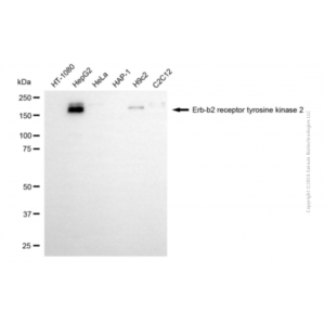
 | Western blotting analysis using anti-erb-b2 receptor tyrosine kinase 2 antibody (Cat#63455). Total cell lysates (30 μg) from various cell lines were loaded and separated by SDS-PAGE. The blot was incubated with anti-erb-b2 receptor tyrosine kinase 2 antibody (Cat#63455, 1:2,500) and HRP-conjugated goat anti-mouse secondary antibody (Cat#101, 1:20,000) respectively. Image was developed using NaQ™ ECL Substrate Kit(Cat#716). |
 | Western blotting analysis using anti-erb-b2 receptor tyrosine kinase 2 antibody (Cat#63455). Erb-b2 receptor tyrosine kinase 2 expression in wild type (WT) and erb-b2 receptor tyrosine kinase 2 (ERBB2) shRNA knockdown (KD) 293T cells with 30 μg of total cell lysates. Hsp90 α serves as a loading control. The blot was incubated with anti-erb-b2 receptor tyrosine kinase 2 antibody (Cat#63455, 1:2,500) and HRP-conjugated goat anti-mouse secondary antibody (Cat#101, 1:20,000) respectively. Image was developed using FeQ™ ECL Substrate Kit (Cat#226). |
 | Flow cytometric analysis of erb-b2 receptor tyrosine kinase 2 expression in HepG2 cells using anti-erb-b2 receptor tyrosine kinase 2 antibody (Cat#63455, 1:2,000). Green, isotype control; red, erb-b2 receptor tyrosine kinase 2. |
 | Immunocytochemical staining of HepG2 cells with anti-Erb-b2 receptor tyrosine kinase 2 antibody (Cat#63455, 1:1,000). Nuclei were stained blue with DAPI; Erb-b2 receptor tyrosine kinase 2 was stained magenta with Alexa Fluor® 647. Images were taken using Leica stellaris 5. Protein abundance based on laser Intensity and Smart Gain:Low. Scale bar, 20 μm. |
You may also be interested in:

