| Reactivity: | Human, Mouse, Rat |
| Applications: | WB, FC |
| Host Species: | Rabbit |
| Isotype: | IgG |
| Clonality: | Monoclonal antibody |
| Gene Name: | flap structure-specific endonuclease 1 |
| Gene Symbol: | FEN1 |
| Synonyms: | MF1; RAD2; FEN-1 |
| Gene ID: | 2237 |
| UniProt ID: | P39748 |
| Clone ID: | 24GB4835 |
| Immunogen: | A synthesized peptide derived from human FEN1 |
| Dilution: | WB 1:1,000-1:5,000; FC 1:2,000 |
| Purification Method: | Affinity purified |
| Concentration: | Lot dependent |
| Buffer: | PBS with 0.02% sodium azide, 50% glycerol, pH7.3. |
| Storage: | Store at -20°C. Avoid freeze/thaw cycles. |
Background
The protein encoded by this gene removes 5' overhanging flaps in DNA repair and processes the 5' ends of Okazaki fragments in lagging strand DNA synthesis. Direct physical interaction between this protein and AP endonuclease 1 during long-patch base excision repair provides coordinated loading of the proteins onto the substrate, thus passing the substrate from one enzyme to another. The protein is a member of the XPG/RAD2 endonuclease family and is one of ten proteins essential for cell-free DNA replication. DNA secondary structure can inhibit flap processing at certain trinucleotide repeats in a length-dependent manner by concealing the 5' end of the flap that is necessary for both binding and cleavage by the protein encoded by this gene. Therefore, secondary structure can deter the protective function of this protein, leading to site-specific trinucleotide expansions.
Images
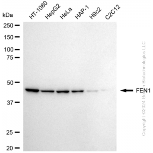
 | Western blotting analysis using anti-FEN1 antibody (Cat#63276). Total cell lysates (30 μg) from various cell lines were loaded and separated by SDS-PAGE. The blot was incubated with anti-FEN1 antibody (Cat#63276, 1:5,000) and HRP-conjugated goat anti-rabbit secondary antibody (Cat#201, 1:20,000) respectively. Image was developed using NaQ™ ECL Substrate Kit (Cat#716). |
 | Western blotting analysis using anti-FEN1 antibody (Cat#63276). FEN1 expression in wild-type (WT) and FEN1 shRNA knockdown (KD) HT-1080 cells with 20 μg of total cell lysates. Hsp90 α serves as a loading control. The blot was incubated with anti-FEN1 antibody (Cat#63276, 1:5,000) and HRP-conjugated goat anti-rabbit secondary antibody (Cat#201, 1:20,000) respectively. Image was developed using NaQ™ ECL Substrate Kit (Cat#716). |
 | Flow cytometric analysis of FEN1 expression in HepG2 cells using anti-FEN1 antibody (Cat#63276, 1:2,000). Green, isotype control; red, FEN1. |
You may also be interested in:

