| Reactivity: | Human, Mouse, Rat |
| Applications: | WB, FC, IC |
| Host Species: | Rabbit |
| Isotype: | IgG |
| Clonality: | Monoclonal antibody |
| Gene Name: | fibroblast growth factor receptor 3 |
| Gene Symbol: | FGFR3 |
| Synonyms: | ACH; CEK2; JTK4; CD333; HSFGFR3EX |
| Gene ID: | 2261 |
| UniProt ID: | P22607 |
| Clone ID: | 25GB2965 |
| Immunogen: | A synthesized peptide derived from human FGFR3 |
| Dilution: | WB 1:1,000-1:5,000; FC 1:2,000; IC 1:100-1:1,000 |
| Purification Method: | Affinity purified |
| Concentration: | Lot dependent |
| Buffer: | PBS with 0.02% sodium azide, 50% glycerol, pH7.3. |
| Storage: | Store at -20°C. Avoid freeze/thaw cycles. |
Background
The gene FGFR3 encodes a member of the fibroblast growth factor receptor (FGFR) family, with its amino acid sequence being highly conserved between members and among divergent species. FGFR family members differ from one another in their ligand affinities and tissue distribution. A full-length representative protein would consist of an extracellular region, composed of three immunoglobulin-like domains, a single hydrophobic membrane-spanning segment and a cytoplasmic tyrosine kinase domain. The extracellular portion of the protein interacts with fibroblast growth factors, setting in motion a cascade of downstream signals, ultimately influencing mitogenesis and differentiation. This particular family member binds acidic and basic fibroblast growth hormone and plays a role in bone development and maintenance. Mutations in this gene lead to craniosynostosis and multiple types of skeletal dysplasia.
Images
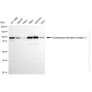
 | Western blotting analysis using anti-fibroblast growth factor receptor 3 antibody (Cat#61593). Total cell lysates (30 μg) from various cell lines were loaded and separated by SDS-PAGE. The blot was incubated with anti-fibroblast growth factor receptor 3 antibody (Cat#61593, 1:5,000) and HRP-conjugated goat anti-rabbit secondary antibody (Cat#201, 1:20,000) respectively. Image was developed using NaQ™ ECL Substrate Kit (Cat#716). |
 | Western blotting analysis using anti-fibroblast growth factor receptor 3 antibody (Cat#61593). Fibroblast growth factor receptor 3 expression in wild-type (WT) and fibroblast growth factor receptor 3 (FGFR3) knockdown (KD) 293T cells with 20 μg of total cell lysates. β-Tubulin serves as a loading control. The blot was incubated with anti-fibroblast growth factor receptor 3 antibody (Cat#61593, 1:5,000) and HRP-conjugated goat anti-rabbit secondary antibody (Cat#201, 1:20,000) respectively. Image was developed using NaQ™ ECL Substrate Kit (Cat#716). |
 | Flow cytometric analysis of Fibroblast growth factor receptor 3 expression in C2C12 cells using anti-Fibroblast growth factor receptor 3 antibody (Cat#61593, 1:2,000). Green, isotype control; red, Fibroblast growth factor receptor 3. |
 | Immunocytochemical staining of C2C12 cells with anti-Fibroblast growth factor receptor 3 antibody (Cat#61593, 1:1,000). Nuclei were stained blue with DAPI; Fibroblast growth factor receptor 3 was stained magenta with Alexa Fluor® 647. Images were taken using Leica stellaris 5. Protein abundance based on laser Intensity and smart gain: High. Scale bar, 20 μm. |
You may also be interested in:

