| Reactivity: | Human, Mouse, Rat |
| Applications: | WB, FC, IC |
| Host Species: | Mouse |
| Isotype: | IgG1 kappa |
| Clonality: | Monoclonal antibody |
| Gene Name: | fms related receptor tyrosine kinase 1 |
| Gene Symbol: | FLT1 |
| Synonyms: | FLT; FLT-1; VEGFR1; VEGFR-1 |
| Gene ID: | 2321 |
| UniProt ID: | P17948 |
| Clone ID: | 24GB7115 |
| Immunogen: | Recombinant protein of human VEGFR1 |
| Dilution: | WB 1:1,000-1:5,000; FC 1:2,000; IC 1:100-1:1,000 |
| Purification Method: | Affinity purified |
| Concentration: | Lot dependent |
| Buffer: | PBS with 0.02% sodium azide, 50% glycerol, pH7.3. |
| Storage: | Store at -20°C. Avoid freeze/thaw cycles. |
Background
This gene FLT1 encodes a member of the vascular endothelial growth factor receptor (VEGFR) family. VEGFR family members are receptor tyrosine kinases (RTKs) which contain an extracellular ligand-binding region with seven immunoglobulin (Ig)-like domains, a transmembrane segment, and a tyrosine kinase (TK) domain within the cytoplasmic domain. This protein binds to VEGFR-A, VEGFR-B and placental growth factor and plays an important role in angiogenesis and vasculogenesis. Expression of this receptor is found in vascular endothelial cells, placental trophoblast cells and peripheral blood monocytes. Multiple transcript variants encoding different isoforms have been found for this gene. Isoforms include a full-length transmembrane receptor isoform and shortened, soluble isoforms. The soluble isoforms are associated with the onset of pre-eclampsia.
Images
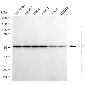
 | Western blotting analysis using anti-FLT1 antibody (Cat#63728). Total cell lysates (30 μg) from various cell lines were loaded and separated by SDS-PAGE. The blot was incubated with anti-FLT1 antibody (Cat#63728, 1:5,000) and HRP-conjugated goat anti-mouse secondary antibody (Cat#101, 1:20,000) respectively. Image was developed using FeQ™ ECL Substrate Kit (Cat#226). FLT1, fms related receptor tyrosine kinase 1. |
 | Western blotting analysis using anti-FLT1 antibody (Cat#63728). FLT1 expression in wild-type (WT) and FLT1 shRNA knockdown (KD) HT-1080 cells with 20 μg of total cell lysates. Hsp90 α serves as a loading control. The blot was incubated with anti-FLT1 antibody (Cat#63728, 1:5,000) and HRP-conjugated goat anti-mouse secondary antibody (Cat#101, 1:20,000) respectively. Image was developed using FeQ™ ECL Substrate Kit (Cat#226). |
 | Flow cytometric analysis of FLT1 expression in HepG2 cells using anti-FLT1 antibody (Cat#63728, 1:2,000). Green, isotype control; red, FLT1. |
 | Immunocytochemical staining of HepG2 cells with anti-FLT1 antibody (Cat#63728, 1:1,000). Nuclei were stained blue with DAPI; FLT1 was stained magenta with Alexa Fluor® 647. Images were taken using Leica stellaris 5. Protein abundance based on laser Intensity and smart gain: High. Scale bar, 20 μm. |
You may also be interested in:

