Sonic Hedgehog (Shh) Rabbit mAb (20 μl)
| Reactivity: | Human, Mouse, Rat |
| Applications: | WB, IF/IC, ELISA |
| Host Species: | Rabbit |
| Isotype: | IgG |
| Clonality: | Monoclonal antibody |
| Gene Name: | Sonic hedgehog signaling molecule |
| Gene Symbol: | SHH |
| Synonyms: | TPT; HHG1; HLP3; HPE3; SMMCI; ShhNC; TPTPS; MCOPCB5; Sonic Hedgehog (Shh) |
| Gene ID: | 6469 |
| UniProt ID: | Q15465 |
| Clone ID: | 8T4D3 |
| Immunogen: | A synthetic peptide corresponding to a sequence within amino acids 200-300 of human Sonic Hedgehog (Shh) (Q15465). |
| Dilution: | WB 1:1000-1:6000; IF/IC 1:100-1:1000 |
| Purification Method: | Affinity purification |
| Concentration: | 0.8 mg/mL |
| Buffer: | PBS with 0.02% sodium azide, 0.05% BSA, 50% glycerol, pH7.3. |
| Storage: | Store at -20°C. Avoid freeze/thaw cycles. |
| Documents: | Manual-SHH antibody |
Background
This gene encodes a protein that is instrumental in patterning the early embryo. It has been implicated as the key inductive signal in patterning of the ventral neural tube, the anterior-posterior limb axis, and the ventral somites. Of three human proteins showing sequence and functional similarity to the sonic hedgehog protein of Drosophila, this protein is the most similar. The protein is made as a precursor that is autocatalytically cleaved; the N-terminal portion is soluble and contains the signalling activity while the C-terminal portion is involved in precursor processing. More importantly, the C-terminal product covalently attaches a cholesterol moiety to the N-terminal product, restricting the N-terminal product to the cell surface and preventing it from freely diffusing throughout the developing embryo. Defects in this protein or in its signalling pathway are a cause of holoprosencephaly (HPE), a disorder in which the developing forebrain fails to correctly separate into right and left hemispheres. HPE is manifested by facial deformities. It is also thought that mutations in this gene or in its signalling pathway may be responsible for VACTERL syndrome, which is characterized by vertebral defects, anal atresia, tracheoesophageal fistula with esophageal atresia, radial and renal dysplasia, cardiac anomalies, and limb abnormalities. Additionally, mutations in a long range enhancer located approximately 1 megabase upstream of this gene disrupt limb patterning and can result in preaxial polydactyly.
Images
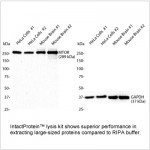
 | Western blot analysis of various lysates using Sonic Hedgehog (Shh) Rabbit mAb (A12695) at 1:1000 dilution. Secondary antibody: HRP-conjugated Goat anti-Rabbit IgG (H+L) (AS014) at 1:10000 dilution. Lysates/proteins: 25μg per lane. Blocking buffer: 3% nonfat dry milk in TBST. Detection: ECL Basic Kit (RM00020). Exposure time: 60s. |
 | Western blot analysis of various lysates using Sonic Hedgehog (Shh) Rabbit mAb (A12695) at 1:1000 dilution. Secondary antibody: HRP-conjugated Goat anti-Rabbit IgG (H+L) (AS014) at 1:10000 dilution. Lysates/proteins: 25μg per lane. Blocking buffer: 3% nonfat dry milk in TBST. Detection: ECL Basic Kit (RM00020). Exposure time: 10s. |
 | Confocal imaging of paraffin-embedded Mouse kidney using Sonic Hedgehog (Shh) Rabbit mAb (A12695, dilution 1:100) followed by a further incubation with Cy3 Goat Anti-Rabbit IgG (H+L) (AS007, dilution 1:500) (Red). DAPI was used for nuclear staining (Blue). Objective: 40x. Perform high pressure antigen retrieval with 0.01 M citrate buffer (pH 6.0) prior to IF staining. |
 | Confocal imaging of Hep G2 cells using Sonic Hedgehog (Shh) Rabbit mAb (A12695, dilution 1:100) followed by a further incubation with Cy3 Goat Anti-Rabbit IgG (H+L) (AS007, dilution 1:500) (Red). The cells were counterstained with α-Tubulin Mouse mAb (AC012, dilution 1:400) followed by incubation with ABflo® 488-conjugated Goat Anti-Mouse IgG (H+L) Ab (AS076, dilution 1:500) (Green). DAPI was used for nuclear staining (Blue). Objective: 100x. |
You may also be interested in:


