Your shopping cart is empty!
")
| Reactivity: | Human, Mouse,Rat |
| Applications: | IHC/IF & ICC/IF |
| Host Species: | Mouse |
| Clonality: | Monoclonal |
| Gene Name: | marker of proliferation Ki-67 |
| Synonyms: | KIA; MIB-; MIB-1; PPP1R105 |
| Gene ID: | 4288 |
| Uniprot ID: | P46013 |
| Immunogen: | KLH conjugated Synthetic peptidecorresponding to Human Ki67 |
| Isotype: | IgG1,κ |
| Purity: | Affinity purification |
| Subcellular location: | Nucleus |
| Documents: | Manual-MKI67 Antibody |
Product Usage Information
IHC/IF | Human, Mouse, Rat | 1: 300-1: 600 | esophageal cancer, ovarian cancer, spleen |
ICC/IF | Human, Mouse, Rat | 1: 100-1: 500 | HeLa |
Background
Enables RNA binding activity and molecular condensate scaffold activity. Involved in chromosome segregation and regulation of mitotic nuclear division. Located in chromosome; nuclear body; and nucleolus. Is active in condensed chromosome. Implicated in several diseases, including Crohn's disease; colorectal cancer; endocrine gland cancer (multiple); graft-versus-host disease; and human immunodeficiency virus infectious disease. Biomarker of several diseases, including Barrett's esophagus; autoimmune disease of musculoskeletal system (multiple); endocrine gland cancer (multiple); gastrointestinal system cancer (multiple); and lung cancer (multiple).
Images
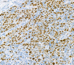
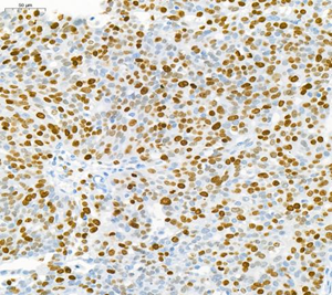
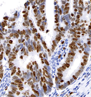
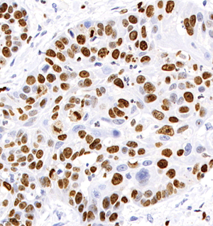
| Immunohistochemistry analysis of paraffin-embedded human esophageal cancer using Ki67 (GB121141) at dilution of 1: 600 |
| Immunohistochemistry analysis of paraffin-embedded human ovarian cancer using Ki67 (GB121141) at dilution of 1: 600 |
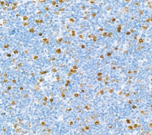
| Immunohistochemistry analysis of paraffin-embedded mouse spleen using Ki67 (GB121141) at dilution of 1: 600 |
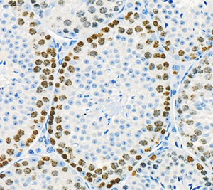
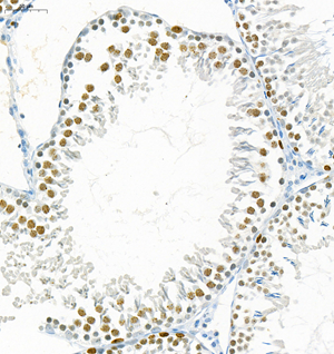
| Immunohistochemistry analysis of paraffin-embedded mouse testis using Ki67 (GB121141) at dilution of 1: 600 |
| Immunohistochemistry analysis of paraffin-embedded rat testis using Ki67 (GB121141) at dilution of 1: 600 |
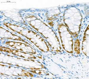
| Immunohistochemistry analysis of paraffin-embedded rat colon using Ki67 (GB121141) at dilution of 1: 600 |
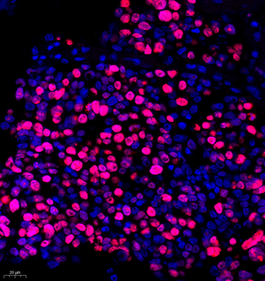
| Immunofluorescent analysis of paraformaldehyde-fixed human colon cancer using Ki67 (GB121141) at dilution of 1: 600 |
| Immunofluorescent analysis of paraformaldehyde-fixed human liver cancer using Ki67 (GB121141) at dilution of 1: 600 |
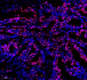
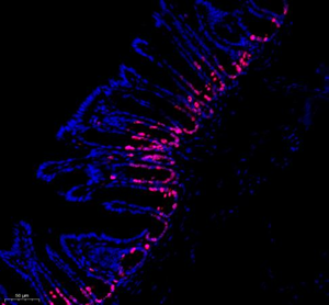
| Immunofluorescent analysis of paraformaldehyde-fixed mouse small intestine using Ki67 (GB121141) at dilution of 1: 600 |
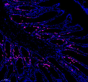
| Immunofluorescent analysis of paraformaldehyde-fixed rat small intestine using Ki67 (GB121141) at dilution of 1: 600 |
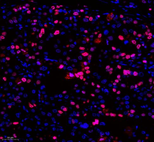
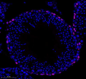
| Immunofluorescent analysis of paraformaldehyde-fixed mouse testis using Ki67 (GB121141) at dilution of 1: 600 |
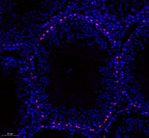
| Immunofluorescent analysis of paraformaldehyde-fixed rat testis using Ki67 (GB121141) at dilution of 1: 600 |
| Immunohistochemistry analysis of paraffin embedded human ovarian cancer using Ki67 (GB121141) at dilution of 1: 500 |
| Immunohistochemistry analysis of paraffin embedded human colorectal carcinoma using Ki67 (GB121141) at dilution of 1: 500 |
| Immunohistochemistry analysis of paraffin embedded human esophageal carcinoma using Ki67 (GB121141) at dilution of 1: 500 |
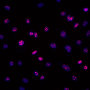
| ICC analysis of Ki67(GB121141)red. Sample: HeLa, 4% PFA (G1101) 20min. Primary antibody: 1: 100, 4°C overnight. Secondary antibody: Cy3 conjugated Goat Anti-Mouse IgG (H+L) (GB21301), 1: 300, RT, 1h. |
| ICC analysis of Ki67(GB121141)red. Sample: NIH-3T3, 4% PFA (G1101) 20min. Primary antibody: 1: 100, 4°C overnight. Secondary antibody: Cy3 conjugated Goat Anti-Mouse IgG (H+L) (GB21301), 1: 300, RT, 1h. |

Storage
| Storage | Store at -20 °C for one year. Avoid repeated freeze/thaw cycles. |
| Storage Buffer | PBS with 0.02%sodium azide,100 μg/ml BSA and 50% glycerol. |
Publications
Wang, F., et al. Lipidomic analysis of plant-derived extracellular vesicles for guidance of potential anti-cancer therapy. Bioact Mater46,82-96(2025). PMID 39737211, IF 18
Sun, L., et al. Dynamic Change of PD-L2 on Circulating Plasma Extracellular Vesicles as a Predictor of Treatment Response in Melanoma Patients Receiving Anti-PD-1 Therapy. J Extra cell Vesicles14,e70054(2025). PMID 40135876, IF 15.5
Yao, H.F., et al.CASC8 activates the pentose phosphate pathway to inhibit disulfidptosis in pancreatic ductal adenocarcinoma though the c-Myc-GLUT1 axis. J Exp Clin Cancer Res44,26(2025). PMID 39865281, IF 11.3
You may also be interested in:

