Your shopping cart is empty!
")
The LiFluor™ 647 EdU Imaging Kit is a state-of-the-art alternative to the classic BrdU cell proliferation assay, expertly optimized for fluorescence microscopy. This assay employs EdU, a modified thymidine analogue, which is seamlessly incorporated into the DNA of newly synthesized cells. It is then labeled with a vibrant, photostable LiFluor™ dye through a rapid and highly-specific click reaction. The fluorescent marking of proliferating cells is both precise and compatible with antibody techniques, a testament to the mild and efficient click protocol utilized.
Key Features
• Simple: Labeling is complete in two steps.
• Efficient: No denaturation steps or harsh treatment required.
• Content-rich results: Better preservation of cell morphology, antigen structure, and DNA integrity.
• Consistent: Not dependent on variable antibody lots for detection.
Specifications
1. Platform: Fluorescence Microscope
2. Detection Method: Fluorescent
3. Ex/Em: 650/665 nm
Applications
Cell proliferation analysis in vitro and in vivo.
Components
1. EdU: 2×1 ml
2. LiFluor 647 azide: 100 µl
3. EdU reaction buffer: 50 ml
4. CuSO4: 1 ml
5. EdU buffer additive: 200 mg
6. Hoechst 33342: 70 µl
Storage
Store at -20°C and protect from light.
Case Study
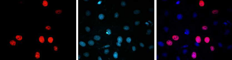
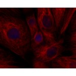
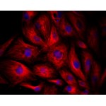
Detection of cell proliferation in cell. A549 cells were treated with 10 µM EdU for 2 hr, then detected with LiFluor™ 647 azide (red), cells were counterstained with DAPI (blue).
Download